


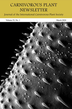
March 2024 CPN
The Carnivorous Plant Newsletter (CPN) is the official publication of the International Carnivorous Plant Society. CPN is published in March, June, September, and December.
Features of CPN include horticultural articles, research papers, field trip reports, conservation news, book and literature reviews, communications from members, cultivar and species descriptions, and meeting announcements.
Contents of Volume 53 Number 1, March 2024
- CPN needs articles from you — Richard Nunn
- Heliamphora electrum (Sarraceniaceae), an enigmatic species of marsh pitcher plant from the Sierra de Lema of Venezuela — Michal R. Golos and Joachim Nerz and François Sockhom Mey and Andreas Wistuba
- Dealing with loss as a horticulturist — Cyrus Exum
- New cultivars — Alessio Ragghianti and Miroslav Srba and Carson Trexler
View the Contents of All CPN Issues
Wed, 04/03/2024 - 16:51
Virtual Events on Zoom
The ICPS is offering Virtual Events on Zoom.
FlytrapStore.com Behind the Scenes Tour
Apr 9, 2024 03:00 PM Eastern Time (US and Canada)
https://us06web.zoom.us/j/5953664257?omn=89900847587
Non-members of the ICPS are welcome.
The Virtual Events are hosted by ICPS Education Director, Kenny Coogan.
Thu, 03/28/2024 - 12:46
ICPS Conference 2024 – Vienna, Austria
The ICPS and GFP (Gesellschaft für Fleischfressende Pflanzen) are excited to announce the GFP will host the next ICPS Conference from 24-26 May 2024 at Orangery of the Schönbrunn Palace in Vienna, Austria.
The Conference will coincide with the GFP 40th anniversary which we would like to celebrate with all of you.
The Orangery of the Schönbrunn Palace is a beautiful venue for this event. There will be a plant sale as well as many lectures on all three days. On Friday evening we will offer a joint evening event and of course there will be the usual conference dinner on Saturday. We will also do our best to offer some excursions after the conference.
Information is available at the conference website
![]()
Fri, 02/02/2024 - 21:58
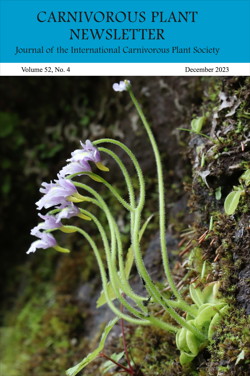
December 2023 CPN
The Carnivorous Plant Newsletter (CPN) is the official publication of the International Carnivorous Plant Society. CPN is published in March, June, September, and December.
Features of CPN include horticultural articles, research papers, field trip reports, conservation news, book and literature reviews, communications from members, cultivar and species descriptions, and meeting announcements.
Contents of Volume 52 Number 4, December 2023
- From the Editors — John Brittnacher
- Erratum — Bob Ziemer
- ICPS Conference 2024 – Vienna, Austria — Christian Dietz and Carsten Paul and Günter Seiter and Daniel Rohrauer
- A new future: Carnivorous plant conservation and the ICPS — Carson Trexler
- Auburn University 2023 Sarracenia oreophila conservation project — Patrick Thompson and Augustus Kirby
- ICPS Conference 2023 – Himeji, Japan — Koji Kondo
- Japanese carnivorous plant groups and organisations — Takaaki Kagawa
- A review of Japanese-native carnivorous plants — Takaaki Kagawa
- The 13th ICPS Post-Conference Tour to the greatest botanical gardens of Hyogo Pref., Japan — Emmi Kurosawa
- The 13th ICPS Post-Conference wetland expeditions — Justin Dunning
- The 13th ICPS Conference Pinguicula ramosa expedition — Emmi Kurosawa
- New cultivars — Emmi Kurosawa
View the Contents of All CPN Issues
Fri, 02/02/2024 - 17:26
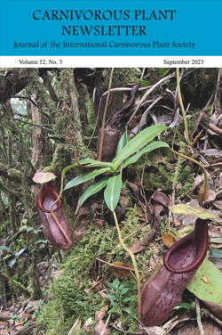
September 2023 CPN
The Carnivorous Plant Newsletter (CPN) is the official publication of the International Carnivorous Plant Society. CPN is published in March, June, September, and December.
Features of CPN include horticultural articles, research papers, field trip reports, conservation news, book and literature reviews, communications from members, cultivar and species descriptions, and meeting announcements.
Contents of Volume 52 Number 3, September 2023
- 13th ICPS International Conference, Japan
- 14th ICPS International Conference, Austria
- Nepenthes limiana (Nepenthaceae), a new pitcher plant from the northern Titiwangsa Range of Peninsular Malaysia — Michal R. Golos and François Sockhom Mey and Andreas Wistuba and Gideon Lim and Stewart R. McPherson and Alastair S. Robinson
- Quick note: Byblis gigantea and B. liniflora traps work the same — Miloslav Studnicka
- Successful cultivation of Darlingtonia californica in a hot climate via root zone chilling — Shawn Lyons
- An inexpensive growth chamber for Heliamphora — Gregory Beylin
- Education Corner — Kenny Coogan
- New cultivars — Mark Rubnitz and Mark S. Anderson and Sergio Mejias Hinojosa and Zihang Qiu
View the Contents of All CPN Issues
Mon, 10/23/2023 - 18:53
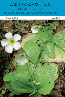
June 2023 CPN
The Carnivorous Plant Newsletter (CPN) is the official publication of the International Carnivorous Plant Society. CPN is published in March, June, September, and December.
Features of CPN include horticultural articles, research papers, field trip reports, conservation news, book and literature reviews, communications from members, cultivar and species descriptions, and meeting announcements.
Contents of Volume 52 Number 2, June 2023
- Support the ICPS Conservation Fund — Carson Trexler
- Pinguicula lithophytica is not conspecific with Pinguicula jaraguana or a variety of Pinguicula jackii — Paul Temple and Ivan Pančo and Cristina M. Panfet Valdés and Yoannis Domínguez
- ICPS Roundtables — Kenny Coogan
- Carnivorous plants and conservation – the role of carnivorous plant enthusiasts — Andreas Fleischmann
- Education Corner: Carnivores in the Classroom update — Kenny Coogan
- New cultivars — Julien Müller and Evan Wang and Emmy Wang and Mark Rubnitz and Arlen Weng and Emanuele Melani and Lukas Mondoux and Michael Richard Sprouse and Fred Vosse and Mentaz Philippe and Guilherme Baião and Bruno Garcia
View the Contents of All CPN Issues
Tue, 07/18/2023 - 18:19